Major Aphthosis and Macrocheilite, Think about Crohn Diseases
ABSTRACT
Orofacial lesion is found in 8-9% of Crohn’s disease (CD) and they are considered as contiguous lesions in the general CD cutaneous disease, we report the case of a young man of 28 years, who presents during a push of his Crohn’s disease a macrocheilitis associated with extensive buccal aphtosis in general CD cutaneous disease
KEYWORDS
Crohn’s disease; Oral granulomatosis; Macrochelitis; Major aphtosis
CLINICAL-IMAGE
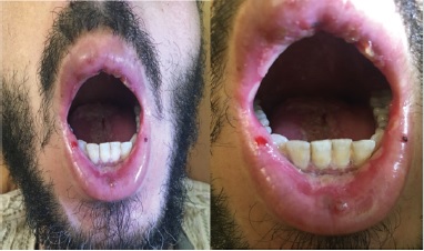
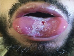
28-year-old patient followed since the age of 22 for digestive Crohn’s disease with poor therapeutic compliance, admitted to the gastrology department for a severe flare-up, a dermatological opinion was sought for lesions of the oral mucosa evolving for 2 months , either 6 weeks before the digestive surge, then gradually worsened. On dermatological examination, a macrocheilitis concerning the upper lip was objectified (Figure 1A) , as well as multiple diffuse aphtosis touching the internal faces of the cheeks, the anterior face of the lower lip and hard palate (Figure 1B), with the presence of an aphthous ulcer of more than 5 cm at the level of the back of the tongue reaching to the buccal floor (Figure 2).
Faced with this aspect, we evoked, specific skin lesions in the context of Crohn’s disease or Behcet’s disease given its frequent association with Crohn’s disease, but we did not have enough criteria to retain this last diagnosis and the patergy test was negative. The histopathological study of a biopsy performed at the level of the macrocheilitis showed a giganto-epithelioid granuloma without caseous necrosis. The diagnosis of a specific oral disease of Crohn’s disease was retained.
During Crohn’s disease (CD), numerous extradigestive localizations have been reported [1]. Dermatological manifestations are described in 32 to 44% of patients with Crohn’s disease [2] In some cases, they appear during known illness while in others, they precede or accompany digestive manifestations, allowing the diagnosis of an intestinal condition that is sometimes clinically latent [3].
The current classification of skin disorders occurring in CD is organized according to the presumed pathologic mechanism. Mucocutaneous lesions are categorized as: CD-specific (involving the skin by a mechanism identical to that occurring in the GI tract); reactive (arising by distinct pathogenetic mechanisms); or associated (noted to occur without a well-defined mechanism) [4]. For specific lesions, the clinical aspects are very varied, and the diagnosis is sometimes based on the discovery of epithelioid and gigantocellular granulomas without caseous necrosis, in the histological study of biopsied fragments [1].
Given this accepted classification, When the granulomatous lesions concern the anoperineal region or the orofacial sphere, these are then contiguous lesions in the general CD cutaneous disease [5]. Orofacial lesion is found in 8-9% of CD [6]. Many aspects can be observed: the prevalence of aphtosis is from 4.2% to 17% [7]. Granulomatous cheilitis is manifested by indurated edema of one or both lips, episodic at first and then permanent. The labial involvement is usually asymmetrical, cracked and is accompanied by a pearlitis. Deep biopsies are necessary to highlight small noncaseous granulomas which allow to retain the diagnosis of orofacial granulomatosis, in the absence of a known intestinal disease, it is always necessary to eliminate a sarcoidosis before retaining the diagnosis of CD [3].
REFERENCES
- Ciubotaru V, Tattevin P, Cartron-Savin L, Le Gall F, Arvieux C, et al. (2003) Cutaneous metastatic manifestations of crohn’s disease. Journal of Internal Medicine 24(3): 198-201.
- Berkowitz EZ, Lebwohl M (2000) Cutaneous manifestations of inflammatory bowel disease. Journal of the European Academy of Dermatology and Venereology 14(5): 349-350.
- Jellali K, Mellouki I, Ibrahimi A (2018) Granulomatosis macro-cheilitis revealing crohn’s disease. Pan African Medical Journal 30: 147.
- Drew JBK, Trevor J, Fangru L, Lisan SP (2014) Metastatic crohn’s disease: A review and approach to therapy. J Am Acad Dermatol 71(4): 804-813.
- Samantha SL, Foster K, Patel D, Shwayder T (2018) Cutaneous manifestations of metastatic crohn’s disease. Pediatr Dermatol 35(5): 566-574.
- Aounallah A, Kahla MB, Ksiaa M, Saidi W, Boussofara L, et al. (2015) Main dermatological manifestations during crohn’s disease: case/control study. Annals of Dermatology and Venereology 144(12): S637.
- Vaillant L, Samimi M (2016) Canker sores and mouth ulcers aphthous ulcers and oral ulcerations. The Medical Press 45(2): 215-226.
Article Type
Clinical Image
Publication history
Received date: January 16, 2020
Published date: January 21, 2020
Address for correspondence
Mounia Bennani, Department of Dermatology, University Hospital Hassan II, Fez, Morocco
Copyright
©2020 Open Access Journal of Biomedical Science, All rights reserved. No part of this content may be reproduced or transmitted in any form or by any means as per the standard guidelines of fair use. Open Access Journal of Biomedical Science is licensed under a Creative Commons Attribution 4.0 International License
How to cite this article
Mounia B, Rhizlane C, Sara E, Zakia Douhi, Hanane B, Fatima Zahra M. Major Aphthosis and Macrocheilite, Think about Crohn Diseases. 2020 - 2(1) OAJBS.ID.000132.